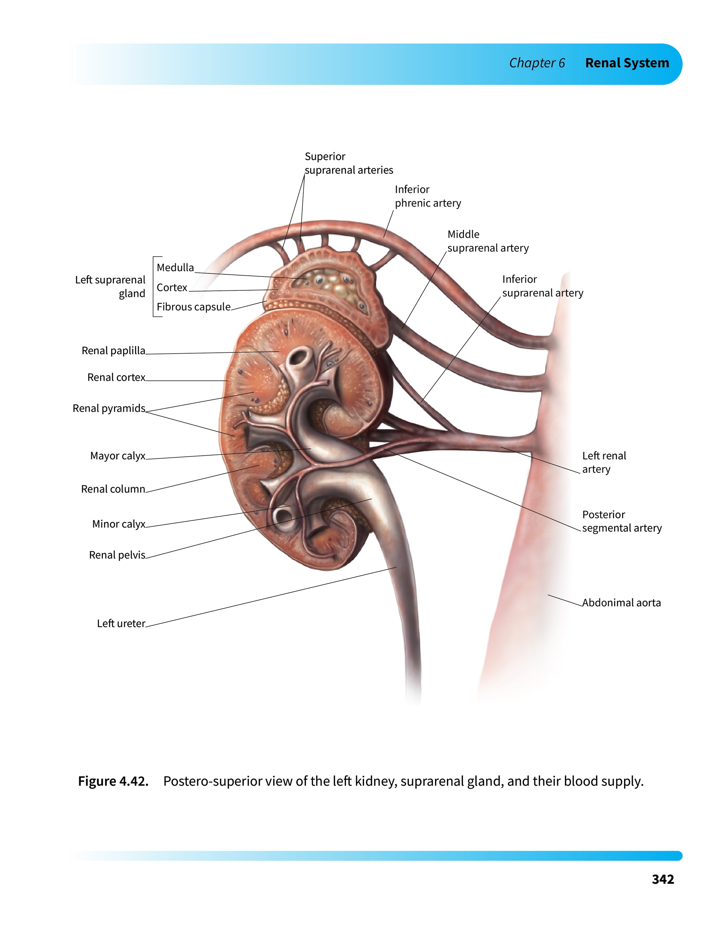
Postero-superior View of the Left Kidney, Suprarenal Gland, and their Blood Supply
‘See The Unsee-able’ Anatomical Illustration
Anatomical Illustration, Medical Illustration
Description
The aim of this project was to visualize an anatomical structure that is impossible to observe directly. As such, this illustration was constructed using a combination of medical imaging data and research.
The posterior view was chosen for this illustration because the sectioned kidney is not well documented from the posterior. As such, I illustrated the way that the posterior segmental artery branches from the abdominal aorta via the renal artery, entering the hilum, and branching. I cut the calyces distally as they transition into the renal pyramids, which allowed me to show their interactions between the posterior segmental artery branches. The three-dimensional nature of the renal pelvis and calyces is often lost in sectional kidney illustrations which segment the calyces proximal or through the renal pelvis. I included a section of the suprarenal gland as illustrations of the internal features of this gland at a gross scale are difficult to find.
Year
2021
Tools
Adobe Photoshop, Adobe Illustrator, Cinema4D, Polymer Clay, Graphite
Media
Print- textbook or anatomical atlas
Client
Michael Corrin
Audience
Students in medical and life science streams and medical professionals
Process
I began by sketching the main anatomy of the kidney based on information obtained from medical imaging data, medical atlases, textbooks, and research papers.
I then obtained a three-dimensional model of a simplified kidney from the Database Center for Life Science, which included the main bulk of the kidney as well as the ureter, renal pelvis, the calyces, and the major anterior arteries and branches that enter the kidney at the hilum. I imported the model in Cinema4D and segmented the bulk of the kidney, leaving the arteries, renal pelvis, and calyces intact. I then modelled additional anatomical features that were missing, such as the posterior segmental artery, the suprarenal gland, the abdominal aorta, as well as the arterial blood supplies between these structures. I then lit and rendered this maquette for use as a drawing reference.
Additionally, a physical maquette was made out of polymer clay (Sculpey) based on direct observation of metal casts of renal pelvises and calyces at Grant’s Anatomy Museum at the University of Toronto to get a better understanding of their three dimensional structures.

SKETCH OF RENAL ANATOMY
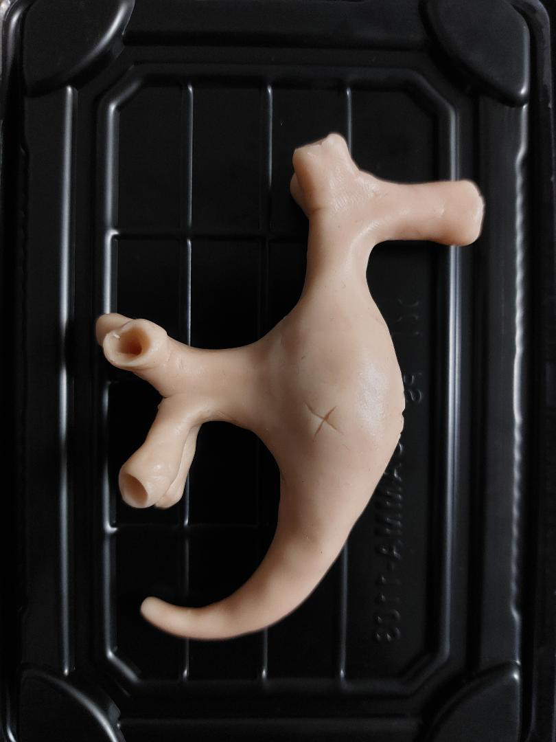
POLYMER CLAY MAQUETTE FROM METAL CASTS OF RENAL PELVISES AND CALYCES AT GRANT’S ANATOMY MUSEUM AT THE UNIVERSITY OF TORONTO

ORIGINAL 3D MODEL OF A KIDNEY FROM THE DATABASE CENTER FOR LIFE SCIENCE
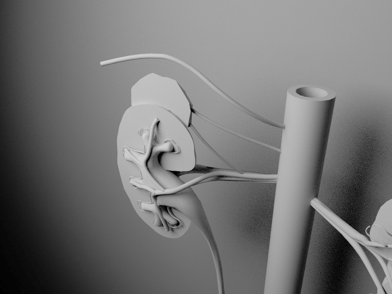
SEGMENTED KIDNEY MODEL WITH ADDITIONAL STRUCTURES MODELLED IN CINEMA4D
Next, the image was rendered in graphite, scanned, and cleaned in Adobe Photoshop.

FINAL SKETCH

GRAPHITE RENDER

DIGITAL RENDERING IN GRAYSCALE
Colour was then added to the grayscale image in Adobe Photoshop.

COLOURED RENDER
Text and graphic design elements were added in Adobe Illustrator.

FINAL ILLUSTRATION
References:
Maquette:
BodyParts3D, © The Database Center for Life Science licensed under CC Attribution-Share Alike 2.1 Japan. https://lifesciencedb.jp/bp3d/
Left kidney; FJ3145_BP49276_FMA7205
Left ureter; FJ3144_BP51070_FMA15572
Anterior division of left renal artery; FJ3458-FJ3463_BP50711_FMA70487
General Structure:
Agur, A. M. R., & Dalley, A. F. (2005). Grant’s atlas of anatomy (11th ed.). Lippincott Williams & Wilkins.
Agur, A. M. R., & Dalley, A. F. (2019). Moore’s essential clinical anatomy (6th ed.). Wolters Kluwer.
Akesson, E. J., Loeb, J. A., & Wilson-Pauwels, L. (1990). Thompson’s core textbook of anatomy (2nd ed.). J. B. Lippincott Company.
Fritsch, H., & Kuehnel, W. (n.d.). Color atlas of human anatomy: Internal organs, vol II (5th ed.). Thieme.
Geddes, L. (2014). Human anatomy: The definitive visual guide. Dorling Kindersley Ltd.
Resnick, M. I., & Parker, M. D. (1982). Surgical anatomy of the kidney. Futura Publishing Company.
Spalteholz, W. (1923). Hand atlas of human anatomy, vol III. J. B. Lippincott Company.
Spence, A. P. (1986). Basic human anatomy (2nd ed.). The Benjamin/Cummings Publishing Company.
Arteries:
Graves, F. T. (1971). The arterial anatomy of the kidney: The basis of surgical technique. John Wright & Sons Ltd.
Ureters/ renal pelvises/ calyces:
Miller, J., Durack, J. C., Sorensen, M. D., Wang, J. H., & Marshall, L. S. (2013). Renal calyceal anatomy characterization with 3- dimensional in vivo computerized tomography imaging. The Journal of Urology, 189(2), 562-567. https://escholarship.org/content/qt1t05q9cp/qt1t05q9cp.pdf?t=nwl3rk
Xu, Z., Li, Z., Guo, M., Bian, H. (2021). Application of three-dimensional visualization fused with ultrasound for percutaneous renal puncture. Scientific Reports, 10.1038/s41598-021-87972-8. https://www.researchgate.net/publication/350970052_Application_of_Three- dimensional_Visualization_Fused_with_Ultrasound_for_Percutaneous_Renal_Puncture
Colour and detail:
Pendovski, L., Vladimir, P., Popovska-Percinic, F., & Ilieski, V. (2021). Silicone plastination procedure for producing thin, semi-transparent tissue slices: A study using the pig kidney. Research Gate. https://www.researchgate.net/publication/267302348_Silicone_Plastination_Procedure_for_Producing_Thin_Semi- _transparent_Tissue_Slices_A_Study_Using_the_Pig_Kidney
Rohen, J. W., Yokochi, C., & Lütjen-Drecoll, E. (1998). Colour atlas of anatomy: A photographic study of the human body (4th ed.). F. K. Schattauer Verlagsgesellschaft mbH, Stuttgart, and Williams & Wilkins.
Standring, S. (Ed.). (2008). Gray’s anatomy: The anatomical basis of clinical practice (14th ed.). Elsevier Ltd.
UT Southwestern Medical Center [UTSWMed]. (2019, May 3). Kidney transplant surgery. Living-donor kidney transplant – 2019 [Video]. YouTube. https://www.youtube.com/watch?v=pke1WhLNoHc
Van De Graff, K. M. (1988). Human anatomy (2nd ed.). Wm. C. Brown Publishers.